Some Examples of Earlier Detection of Breast Cancer
02.03.98. Mammography - fibrocystitis mastopathy, fibrous tissue prevails. Scar changes.
02.03.98. CBE - the both breasts with diffuse fibrocystitis mastopathy. The operation scar is solid at some parts. Nodes are not detected. The region lymph nodes are not enlarged. Oncologist diagnosis - diffuse fibrocystitis mastopathy.
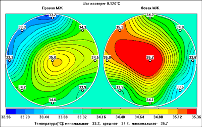
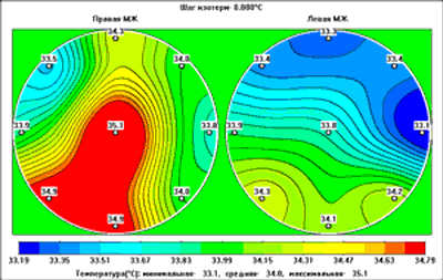
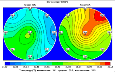
04.03.98. RTM-Diagnosis - the thermogram has features of left breast cancer locating in the upper inner quadrant.
02.03.98. Biopsy - Erythrocytes. Fat drops.
Considering RTM-Diagnosis a control examination was ordered in 3 months.
In 4 months.
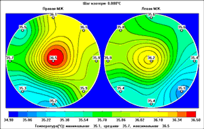
08.07.98. RTM-Diagnosis - the thermogram has features of left breast cancer locating in the upper inner quadrant.
08.07.98. CBE - The left breast has scar changes. Under the areola there is a solid elastic lump of 1 cm in diameter with an enough exact boundary. The region lymph nodes are not enlarged. Oncologist diagnosis: nodal fibrocystitis mastopathy, breast cancer suspicions.
14.07.98. Biopsy - unstructured masses, isolated leucocytes.
16.07.98. Biopsy - a cancer cytogram.
16.07.98. Mammography - Fibrocystitis mastopathy, in the fibrous tissue there is a lump of 1.5 cm in diameter with a light boundary.
Final oncologist diagnosis: left breast cancer.
22.09.97. Mammography - preclimacteric menstrual cycle disorders. Palpation: in the right breast in the upper inner quadrant there is an elastic lump. X-ray: the both breasts with diffuse fibrocystitis mastopathy. Glandular tissue is more solid in the inner quadrants of the right breast. Diagnosis: right breast cyst - ?.
22.09.97. CBE - complaints on solid neoplasm in the right breast from February, 1997. Gynecology - healthy, menstruation is irregular, the premenopause period, Р-1, А-5, В- 0. In April, 1997 the gall-bladder was ablated. Objective: the breast is developed properly. The nipples, areolas are normal. There is not nipple discharge. On the background of moderate fibrocystitis mastopathy in the right breast in the upper inner quadrant there is a solid elastic lump of 3.5 - 4 cm in diameter without an exact boundary. Skin symptoms are absent. The regional lymph nodes are normal. Diagnosis: nodal fibrocystitis mastopathy. Cancer suspicions?
30.09.97. RTM-Diagnosis - the thermogram has features of breast cancer located in the upper inner quadrant.
22.09.97 - 30.09.97. Biopsy - a glandular solid cancer cytogram.
30.09.97. Final diagnosis: right breast cancer.
16.01.97. Mammography – on the border of outer quadrants in the left breast there is a irregular lump of 3.5 cm in diameter. The breast structure is changed diffusely. The skin and areola are incrassate. Diagnosis: infiltrating cancer of the left breast.
16.01.97. CBE – complaints on a painful lump in the left breast. It was noted by patients two days ago. The temperature was 38?С.
Objective – the left breast has serious hyperemia, it is inflammative, the nipple is retracted. On the border of the outer quadrants there is a large infiltrate, palpation is painful. In the left axillary region an enlarged lymph node is palpated. Diagnosis: acute mastitis.
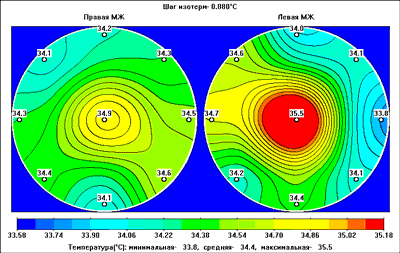
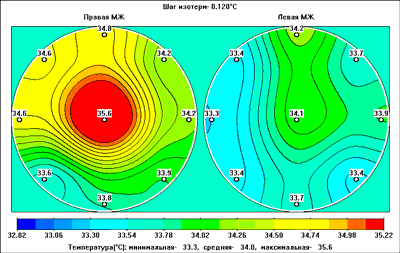
16.01.97. RTM-Diagnosis - the thermogram has features of left breast cancer.
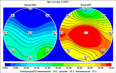
In five months the patient had repeated RTM-Diagnosis.
03.07.97. RTM-Diagnosis - the thermogram has features of left breast cancer located in the lower outer quadrant.
In this case (Patient P, 58 year old) there are positive changes resulted by the treatment of acute mastitis, however diagnosis is not changed - the thermogram has features of left breast cancer.
In 1997 the patient complained on strong pain in the left breast, in August, 1997 she was operated as a result of chronic mastitis. Plan histology detected ductal cancer.
09.12.97. Mammography - Complaints on hardening and pain in nipple.
BE - a solid lump in the right areola close-fitting to it.
X-ray - diffuse fibrocystitis mastopathy. The structure of the lump can not be detected as a result of glandular triangle. The patient is directed to an oncologist, biopsy guided by palpation in order to exclude the existing process.
10.12.97. CBE - complaints on a solid mass under the right nipple close fitting to the nipple. Objective: there is a solid glandular lump under the right nipple. Diagnosis: nodal fibrocystitis mastopathy of the right breast. (R - Susp.)
10.12.97 - 17.12.97. Biopsy - № 1- there are destroyed cells in erythrocyte groups. № 2 - erythrocytes, separated cubic epithelium cells.
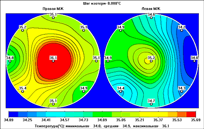
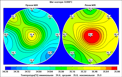
17.12.97. RTM-Diagnosis - the thermogram has features of breast cancer.
09.01.98. Final oncologist diagnosis: nodal fibrocystitis mastopathy of the right breast. (R - Susp.)
Extract from the case history #7401.
Patient Z., 49 year old, was treated in the Oncology Surgery Department of the Ostroumov Municipal Hospital #33 from 16 March to 6 April 1998. She was diagnosed with right breast cancer, 1st., T1N0M0.
The diagnose was made after plan histology after central resection performed on 3 March 1998. The Patient was hospitalized repeatedly.
20 March 1998 the patient had radical Paity mastectomy of the right breast.
Histology #16813-822 and 22086-105: infiltrative lobular breast cancer of less than 0.5 cm in diameter.
3.10.97. Mammography - on the background of tissue remainders in the left breast, in the upper outer quadrant there is a lump with small and micro-calcinators of 3.8x3.5 cm. Diagnosis: nodal mastopathy with calcinators, cancer suspicions.
3.10.97. CBE - complaints on a painful lump in the left breast. It was noted a month ago and since then is has been increasing. Objective: the beasts are small, the nipples and areolas are normal. Moderate fibrocystitis mastopathy. A solid lump of 1.5 cm without an exact boundary is palpated in the upper outer quadrant. In the upper outer quadrant a movable lump of 3 cm without an exact boundary is palpated. There are not any skin symptoms. In the left axillary region there is a round solid lymph node of 1.5 cm. Diagnosis: Cr mam.sin. IIIb T3N1M0.
3.10.97. Cytology - 1) Erythrocytes, lymphocytes, destructurally changed elements, stromal elements of the fibroplast type. Cancer elements are not detected. 2) Erythrocytes, destructural mass, proliferative cubic epithelium cells in which there are some large cells that may be cancer cells. The lymph node – erythrocytes, lymphocytes of different age. Cancer elements are not detected.
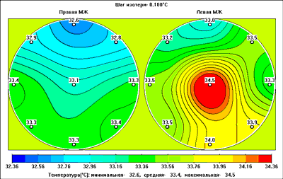
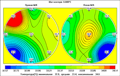
3.10.97. RTM-Diagnosis - There are not features of breast cancer.

3.04.98. Mammography - on the background of bilateral diffuse fibrocystitis mastopathy on the border of the left upper quadrants there is a part of many micro-calcinators with a lane to the nipple. The diameter of the part is ? 3.5 cm. The previous mammogram was not shown. Diagnosis: Neo mam.sin.
CBE - left breast cancer T3N1M0 (dynamics is absent).
28.02.2000. Mammography - fibrofat involution, fibrous component in the both breasts. There are small and micro-calcinators in the left breast.
28.02.2000. Consultation with a professor - in mammograms in the left breast there are many macrocalcinators in the area of 6x3 cm. Nodes are not detected. Skin is normal. Palpation – micromastopathy, nodes are not detected, the lymph nodes - abs, there is not nipple discharge.
Final diagnosis: moderate fibrocystitis mastopathy.
24.12.97. Mammography - bilateral fat involution, nodes are not detected. A fibrous lane from the left indrawn nipple.
24.12.97. CBE - the left nipple was indraw for 2 year, as the patient said.
Gynecology - mioma - 1971г. Objective: the left nipple is indrawn. It can not be pull out, there is a lump without an exact boundary on the border of the outer quadrants. The regional lymph nodes are normal. Diagnosis: ductal left cancer suspicions.
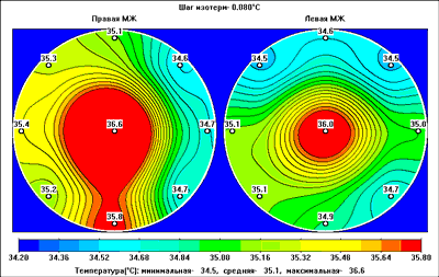
24.12.97. RTM-Diagnosis - the thermogram has features of left breast cancer.
24.12.97. - 14.01.98. Biopsy - erythrocytes, fat drops.
26.02.98. Final oncologist diagnosis - Susp. bl. mam. sin. (ductal cancer), the patient was hospitalized to the Municipal Hospital #62 for sector resection.
08.04.98. In the Municipal Hospital #62. Diagnosis - left breast cancer; radical mastectomy of left breast was performed.
29.05.98. Mammography - in the left breast on the border of the outer quadrants there is a lump of 2.5x1.5 cm with an even, at some parts inexact boundary. Diagnosis: localized fibrocystitis mastopathy? Cyst?
29.05.98. CBE - complaints on pain in the breasts for two weeks, a lump in the left breast. Objective - fibrocystitis mastopathy of the breasts. In the left breast on the border of the outer quadrants there is a solid, palpated lump of 3x4 cm with an enough exact boundary. There are not skin symptoms. The regional lymph nodes are not enlarged. Diagnosis: Nodal fibrocystitis mastopathy.
29.05.98. RTM-Diagnosis - the thermogram has features of breast cancer located on the border of the outer quadrants.

29.05.98- 2.06.98. Biopsy - cytologycally - proliferative mastopathy, cancer suspicions.
02.06.98. Final oncologist diagnosis: left breast cancer suspicions.
19.01.98. Mammography - on the background of bilateral fibrocystitis mastopathy in the right breast on the border of the outer quadrants there is an oval lump of 3.0 in diameter with an inexact boundary at some parts. Diagnosis: a cyst in the right breast, cancer suspicions.
19.01.98. CBE - complaints on a solid mass in the right breast, noted in September 1998. Gynecology - healthy. Objective - in the right breast near the areola, on the border of the outer quadrants there is a solid elastic lump of 3 cm in diameter. Skin symptoms are absent. The regional lymph nodes are normal. Diagnosis: nodal fibrocystitis mastopathy.
23.01.98. RTM-Diagnosis - the thermogram has features of right breast cancer located in the lower outer quadrant.
19.01.98 - 29.01.98. Biopsy - a cancer cytogram.
23.01.98. Repeated mammography - in guided and side mammograms there is a lump without an exact boundary. Diagnosis: right breast cancer suspicions.
Final oncologist diagnosis: right breast cancer, IIb T2N0M0.
10.12.97. Mammography - bilateral diffuse fibrocystitis mastopathy. R-control - November 1998.
27.02.98. CBE - solid breast. Nodes are not detected. Diagnosis: more intensive diffuse fibrocystitis mastopathy in the right breast.
27.02.98. CBE - complaints on pain in the right breast. Gynecology - menstrual cycle disorders. Objective – in the right breast in the lower outer quadrant near the areola the skin is retracted for several years. Bilateral fibrocystitis mastopathy. In the right breast, where the skin is indrawn more solid mass without an exact boundary is palpated. Diagnosis: right breast cancer suspicions.
27.02.98. RTM-Diagnosis - the thermogram has features of right breast cancer. The examination was conducted on an incorrect menstrual cycle day.
27.02.98 -3.03.98. Biopsy - erythrocytes, gemosiderofages, separated cubic epithelium cell groups, complexes of polimorphous cancer cells.
03.03.98. Repeated mammography - in the pictures guided to the palpated lump there are two parts of nodal mastopathy of 1.5 cm with inexact boundaries. Right breast cancer suspicions.
Final oncologist diagnosis: right breast cancer.
Ultrasound - Bilateral diffuse mastopathy, in the left breast in the upper outer quadrant there is a part of high geterogeneity with a cyst of 5 mm in diameter (the square of the area is 14x10 mm) - mastopathy changes, other pathologies are not detected.
Mammography – in the left breast on the background of diffuse fibrocystitis mastopathy on the border of inner quadrants there is a micro-calcinator group and there are two groups near the areola. Diagnosis: left breast cancer suspicions.
RTM-Diagnosis - the thermogram has features of left breast cancer.
Biopsy - cancer with cricoid cells.
Preliminary diagnosis - bilateral mastopathy.
Final diagnosis: left breast cancer, IIA T2N0M0.
The patient had subtotal radical resection of the left breast with plastics and radiotherapy.
Histology - K-15867-91: Invasive ductal cancer, ductal component prevails, II stage (6 balls).
Postoperative diagnosis: left breast cancer IIA T2N0M0.
RTM-Diagnosis Protocol№ 0014АА00001А.
Mammography – in the right breast in the upper outer quadrant there is a fibroadenoma of 2.5 cm with an inexact boundary. Diagnosis: right breast cancer suspicions.
RTM-Diagnosis - the thermogram has features of breast cancer located in the upper outer quadrant.
Biopsy - cancer with mucus.
Final diagnosis: right breast cancer, IIb T2N1M0.
RTM-Diagnosis Protocol № 0001АА01175А.
6.07.98. Mammography - on the background of fibrocystitis mastopathy in the left breast in the upper inner quadrant there is an irregular lump without an exact boundary. Diagnosis: left breast cancer suspicions.
6.07.98. CBE - gynecology - myoma of the uterus for 17 weeks. Objective: in the left breast in the upper inner quadrant there is a solid elastic lump of 2 cm in diameter with an enough exact boundary. Diagnosis: cyst (Rg.- Susp.BL.mam.sin).
10.07.98. RTM-Diagnosis - the thermogram has features of left breast cancer located in the upper inner quadrant.
10.07.98. Biopsy - a cancer cytogram.
Final oncologist diagnosis: left breast cancer, T1N0M0 Ib stage.
Mammography - on the fibrous background in the right breast there is a lump. Diagnosis: nodal fibrocystitis mastopathy, right breast cancer suspicions.
CBE - in the right breast in the upper outer quadrant there is a lump of 2.5 cm in diameter. Diagnosis: nodal fibrocystitis mastopathy, right breast cancer suspicions.
RTM-Diagnosis - the thermogram has features of right breast cancer located on the border of the upper quadrants.
Biopsy - fat drops.
Repeated biopsy - proliferative mastopathy.
Postoperative diagnosis: right breast cancer, T1N1M0 Ia stage
www.resltd.ru © 2001 RES, Ltd. Created and Designed by V.I.